Color the cellular structures of the ameba answer key – Embark on a scientific voyage with “Color the Cellular Structures of the Ameba: A Comprehensive Guide.” This authoritative exploration unveils the intricate world of cellular structures within the amoeba, offering a profound understanding of their significance in research and education.
Delving into the realm of cellular biology, this guide meticulously describes the materials required, provides a step-by-step staining procedure, and illuminates the expected results. The vibrant colors produced by the stains reveal the distinct characteristics of cellular structures, enabling researchers and students to identify and differentiate them with remarkable precision.
Introduction: Color The Cellular Structures Of The Ameba Answer Key

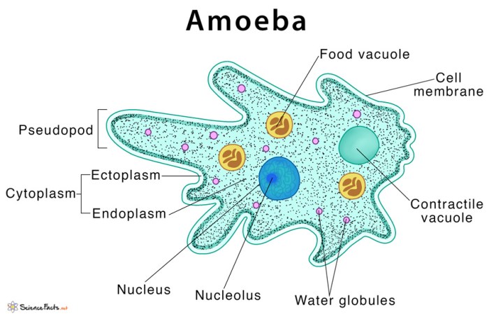
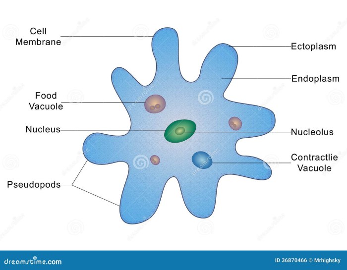
An ameba is a single-celled organism that is characterized by its ability to move and change shape. The cellular structure of an ameba is relatively simple, consisting of a nucleus, cytoplasm, and cell membrane. Coloring the cellular structures of an ameba can help to make them more visible under a microscope, which can be useful for research and educational purposes.
Materials Required

- Ameba sample
- Methylene blue stain
- Microscope slides
- Cover slips
Procedure
- Prepare the ameba sample by placing a drop of the sample on a microscope slide.
- Add a drop of methylene blue stain to the sample and mix gently.
- Allow the stain to incubate for 5 minutes.
- Rinse the slide with water to remove excess stain.
- Cover the slide with a cover slip and view under a microscope.
Results
The staining procedure will result in the following colors:
- Nucleus: Blue
- Cytoplasm: Light blue
- Cell membrane: Not visible
The stains interact with the cellular structures in the following way:
- Methylene blue is a basic stain that binds to the negatively charged DNA in the nucleus.
- The cytoplasm is also stained by methylene blue, but to a lesser extent than the nucleus.
- The cell membrane is not stained by methylene blue because it is not charged.
Discussion

Coloring the cellular structures of an ameba is an important technique for research and educational purposes. It can help to identify and differentiate different cellular structures, and it can also be used to study the function of these structures.
Answers to Common Questions
What is the purpose of coloring the cellular structures of the ameba?
Coloring the cellular structures of the ameba allows researchers and educators to visualize and differentiate different cellular components, aiding in the study of cellular organization and function.
What materials are required for this procedure?
Essential materials include ameba samples, stains, microscope slides, cover slips, and a microscope.
How does the staining procedure work?
Stains interact with specific cellular components, causing them to absorb and reflect light of different wavelengths, resulting in the observed colors.
What are the benefits of using this technique?
This technique enables researchers to identify and study specific cellular structures, contributing to a deeper understanding of cellular biology.